Cell toxicity / Proliferation
Apoptosis Assay
Cell cycle analysis

cell invation / migration assay
Disease model
Phagocytic assay
Custom Assays

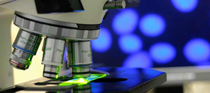
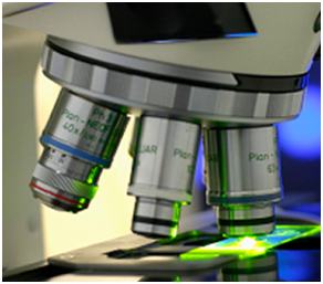
Cell-based assays, bioassay and their development can range from cell proliferation, cytotoxicity assays to cell metabolic and cell-based immuno-assays. Mechanisms of action of a drug candidate can be further investigated by looking at receptor binding/activation, cell signaling, drug internalization and subcellular localization and movement of critical proteins/analytes. Our expertise in in vitro cell-based and cell-free assays provide valuable tools for your drug discovery approaches.
At MuriGenics, for most applications, we provide high-throughput screening assays. We have optimized protocols using 96-well plate format supported by our multi-mode spectrophotometer multi-cytokine Multiplex analyzer, multi-mode and real-time cell imager (Brightfield, color, and fluorescence) and flow cytometer. Our instrumentation supports fluorescence, luminescence, and colorimetric readouts as well as kinetic, end point and real time cell morphology information. With our expertise, we can help you develop high-throughput screening assays for your drug candidates.
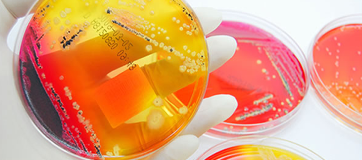
We have over 200 banked cell lines; cell line panels for your research. These cell line panels can be categorized by cancer type, e.g.
- Breast- 4T1, EMT-6
- Lymphoma- A20, EG7.ova
- Colon- Colon 26, CT26, MC38
- Melanoma- B16F10
- Renal- Renca
- Lung- KLN205, Lewis lung
Primary cell culture
Isolation/propagation of cells from tissues (e.g. T-cell isolation from Spleens, PBMCs for cell activation/proliferation study, Fibroblast from Skin etc.).
Cell culture optimization
We can optimize growth of modified and unmodified cell lines using matrices, serum-free media, 3D cell culture etc. for your research needs.
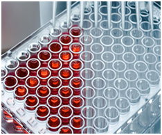
Cell toxicity / proliferation assays
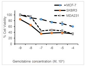
Your drug candidate may be killing, inhibiting or enhancing the cell proliferation. A 96-well format cell viability assays can be performed to investigate this. We have over 200 banked cell lines from various cancers which can be used for your research. Our routine cell viability assay uses cell-permeant compound that is rapidly taken up by cells. The reducing environment within viable cells converts the reagent to an intensely fluorescent dye which can be detected by measuring fluorescence or absorbance. We can also adopt MTT or ATP based luminescence assay to quantify cell viability. IC50 or EC50 curves can be generated and potency of the drug candidates on multiple cell lines can be evaluated. We have cell-line panels for breast, lung, colon, melanoma, leukemia and glial cell types which can aid your screening approach. These
Cell Death / Apoptosis assay
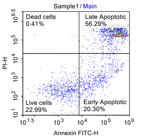
Apoptosis is characterized by certain morphological feature including loss of plasma membrane asymmetry and integrity. In the apoptotic cell, the membrane phospholipid phophatidylserine (PS) is translocation from the inner to outer leaflet of the plasma member there by exposing PS to the external cellular environment. Annexin V, a phospholipid binding protein can bind to PS with high affinity and Annexin V conjugated to a fluorophore can be deterred by flow cytometry. Externalization of PS occurs at an early stage of apoptosis, thus Annexin based assay can be detect early apoptotic cell in your cell death/toxicity assay. Dead cells can be separately detected by flow cytometry following staining with propidium iodide (PI) or 7-amino actinomyciin staining (7-AAD).
Cell cycle analysis
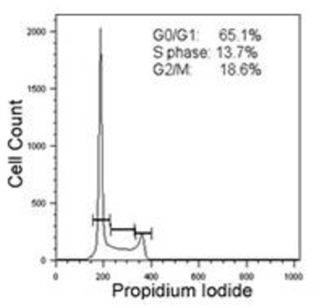
The inhibition of the cancer cell growth by your test compound can be associated with cell cycle arrest at a particular phase of cell cycle. Since DNA doubles during the S phase, the DNA content of the cells can detected using fluorescent DNA binding dyes and flow cytometry. Flow cytometry can distinguish cells in different phase of the cells cycle depending on their cellular DNA content. Cellular DNA content frequency histograms can yield information about the distribution of cells in the three major phases of the cell cycle (G1 vs S vs G2/M). The DNA content in turn can be tied to cyclin D, E, A or B1 expression, thus delineating G0 from G1 cells.
At MuriGenics, PI or BrDU staining can be used for DNA staining and Ki-67 immunostaining can also be explored. Such cell cycle analysis can help you understand the mechanism of action of your drug candidate
Cell migration and invasion assay
In normal physiology and immune-oncology, cell migration/invasion is a complex multi component process that plays a vital role in the progression of various diseases including cancer, autoimmune disease and atherosclerosis. Quantitative determination of Cell migration/invasion also include degradation of extra cellular matrix (ECM) component by proteolysis.
We provide assays for the quantitative determination of various factors on cell migration, adhesion and invasion. These include assays for investigation of pharmacological agents, evaluation of adhesion molecules, receptors, chemoattractant or other molecules responsible for cell migration.
Cell Migration assay
This assay allows appropriate and sensitive quantification of cell migration towards a chemical concentration gradient (chemotaxis).
Cell Invasion assay
This assay allows appropriate and sensitive quantification of cell invasion through collagen, fibronectin and basement membrane ECM matrix. This assay can also be adapted to have a layer of cells such as endothelial cells.
Our assays include –
I) Transwell chamber assays (Boyden chamber) allows for the investigation of transmigration of immune or cancer cells from the inserts pores based on the presence/absence of chemoattractant stimuli. Your drug candidate may influence the transmigration/invasion and may be useful for controlling metastasis or autoimmune response. The transmigration/invasion barrier can be the membrane insert itself of different pore sizes, endothelial cells, ECM matrix, collagen, fibrinogen coated inserts.
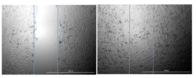
II) Scratch assay allows migration of cells to the site of scratch on the plate/chamber slide. The migration is monitored over a period of time in the presence or absence of drug compound. Finding drugs that can inhibit migration of immune cells to the site of autoimmune site thus provides an appealing approach to alleviate the severity of autoimmune disease.
In vitro Disease models
Several disease models can be partially replicated in cell models. We can perform pathway driven investigation in the available cell lines.
HepG2, the liver carcinoma cell line provide a cell line model for nonalcoholic steatosis hepatitis model where stress molecules are upregulated which can be detected by quantitative PCR.
Drug-induced hepatic injury is a common reason for the withdrawal of an approved drug. Lists of assessment that MuriGenics can perform includes – CYP inhibition/induction (drug-drug interaction), LDH leakage, pathology specific hepatotoxicity (steatosis, NASH etc..), transporter assay (ATPase etc.) to aid our clients research.
Drug-induced hepatotoxicity ranges from asymptomatic elevation of liver enzymes to fulminant hepatic failure. We perform a series of assay screens that may be utilized throughout the discovery and development phases in conjunction with traditional in vivo methods to aid the client in the selection of a lead compound. Below is a list of assessments that we perform:
- Morphological Evaluation
- LDH Leakage
- MTS
- Pathology Specific Hepatotoxicity
- Steatosis
- Phospholipidosis
- CYP Inhibition/Induction (Drug/Drug Interactions)
- Cytokine analysis ATPase Assay (Transporters)
- Morphological Evaluation
- LDH Leakage
- MTS
- Pathology Specific Hepatotoxicity
- Steatosis
- Phospholipidosis
- CYP Inhibition/Induction (Drug/Drug Interactions)
- Cytokine analysis ATPase Assay (Transporters)
Phagocytosis Assay
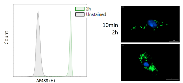
The Phagocytosis Assay provides a way for researchers to observe and quantitate the process of phagocytosis polynuclear cells and mouse macrophages by following the internalization of a foreign particle labeled with the fluorescent dye. The methodology we use has been developed using an adherent murine cells; however, by modifying the cell culture conditions, researchers can adapt this phagocytosis assay to other adherent cell lines.
We also offer to label(insert link here) your peptide/protein of interest with a fluorescent dye and follow the lablel qualitatively and quantitatively during the pagocytosis process.
Custom Assays
We can also consult with you to design the assay you want. We have in the past done cell in assays covering cell signaling, subcellular localization and drug internalization.