Basic H&E
Immunohistochemistry / Immunofluorescence
Tissue array or tissue microarray (TMA)

Multiplex Immunohistochemistry

Biomarker staining development
Histology is the microscopic study of cells and tissues. The specimen is studied after having been sectioned, stained, and mounted on a microscope slide. At MuriGenics, we have extensive expertise in providing high-quality histologic sections on a wide selection of specimens to support your research. Our services include tissue fixation, procession, sectioning, staining, immunohistochemistry, in situ hybridization and method development.
Basic histology
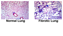
To aid your in vivo studies, routine paraffin and frozen section preparation (FFPE) followed by hematoxylin and eiosin staining is available along with the wide selection of histochemical stains – H & E, Masson’s Trichrome, Toluidine Blue, Sirius Red, LFB, PAS, Luna etc..
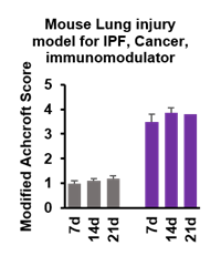
Specialized histological methods are also available. Our experts can assist you with special procedures and project design when requested. We also have capability to do histopathology and report important anatomical pathology. Establishment of a disease model and its treatment by your investigational compound can be assess by scoring the pathology of relevant tissue/cell type.
Our highly trained and experienced board-certified pathologist is committed to provide you with the highest level of quality. Our experienced and detail-oriented technical personnel work alongside with our pathologist, in vivo staff and study director/s.
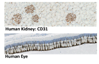
Immunohistochemistry / Immunofluorescence
We are able to customize an immunohistochemical assay for your particular research and development needs and/or to complement any of our disease model studies. Our services include screening antibodies as well as optimizing our assays to ensure highly specific and robust expression detection. We can develop and adopt best methodology to detect your antigen in PPFE and cryopreserved samples.
Tissue array or tissue microarray (TMA)
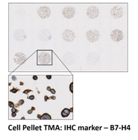
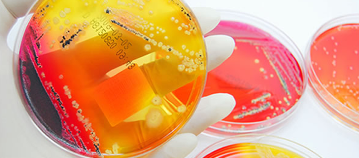
Tissue microarray allows for high throughput platforms for the rapid analysis of molecular markers associated with disease diagnosis and therapy. TMA can be used to validate the clinical importance of novel biological targets in the development of therapies, study of new protein markers and diagnostics.
MuriGenics offer study designs to incorporate TMA studies with your research and development project. Therapeutic antibodies can be tested before FDA approval with multiple organ tissue array. Cancer array with stage, grade and control tissues can also be run.
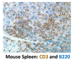
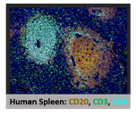
Multiplex Immunohistochemistry
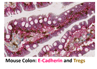
We, at MuriGenics can use multiplex immunohistochemistry technique to simultaneously investigate the expression pattern of multiple protein involvement in pathways of interest within the context of preserved tissue architecture.
Immune check point markers can be co-detected and spatially characterize with mIHC. Our mIHC can simultaneously detect multiple (~3) targets of interest.
Biomarker staining development
Various histological stains can be used to enhance the visualization of different cells and is an essential tool of biology and medicine. We specialize in using optimized methods to best preserve the tissue for exacting cellular details for your study.
Challenging staining protocols can be optimized at MuriGenics with development project. We have access to –
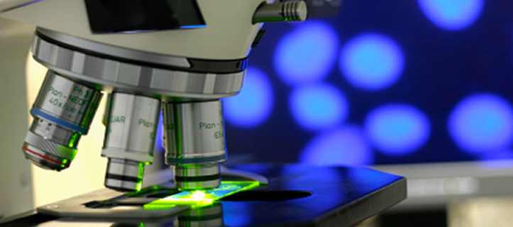
- Laser Scanning Confocal Microscopy (LSM)
- Automated slide scanner for fluorescence or brightfield
- Multi-mode image applications including immunofluorescence and histology using Citation 5 (Biotek). It has the ability to collect phenotypic and quantitative data